Mid-aged mice treated for 20 weeks with nicotinamide mononucleotide (NMN) – a precursor to a vital molecule – show improved fertility potential and reduced aging of ovaries.
By Jonathan D. Grinstein, Ph.D.
Highlights
The relationship between female fertility and aging is complex. We know for sure that the number of eggs (ovum) and the window for their fertilization are limited by ovarian aging. As women get older, the ability to support conception shrinks like a reverse snowball: the quantity and quality of structures that can support the ovum called ovarian follicles gradually decline, ending with menopause. Since menopause and the preceding decline in oocyte quality seem to have a fixed time interval, treatments that expand the female reproductive lifespan are highly sought after.
Researchers from Jiangsu University in China demonstrate that long-term treatment with nicotinamide mononucleotide (NMN) has anti-aging effects on ovaries. In an article published in The Journal of Nutritional Biochemistry, Huang and colleagues show that 20 weeks of oral NMN treatment (0.5 mg/mL/day) could improve mid-aged ovary aging, preserving the ovarian reserve. In mid-aged mice, this NMN treatment regimen improved the structure of ovaries and reduced senescence — a phenomenon characterized by the cessation of cell replication, which is linked to aging.
“Our work expounds a theoretical basis for application of NMN to improve the fertility of aged women,” said Huang and colleagues.
The menstrual cycle for dummies
A major aspect of reproductive capacity in women is its cyclical activity, a feature strikingly reflected in the growth and development of dominant follicles. Ovarian follicles are tiny sacs filled with fluid that secrete hormones to influence menstrual cycle stages and have the potential to release an egg (ovum) for fertilization.
Typically, the human ovaries produce a single dominant follicle that results in each menstrual cycle’s single ovulation. These reproductive structures progress through several distinct phases before it releases an ovum, starting from a reserve of quiescent primordial follicles set up in early life and ending with either ovulation or follicular death.
During each menstrual cycle, several follicles develop in the ovaries. Each follicle contains an egg, but only one follicle continues to develop. Next, this follicle releases an egg (ovulation), and the ruptured follicle closes after releasing the egg and forms a corpus luteum. If the egg is not fertilized, the corpus luteum degenerates, and, depending on ovarian age, a new menstrual cycle begins.
Phases of the human menstrual cycle.
The normal human menstrual cycle is 28 days. On this basis the cycle is described as starting with about five days of menstruation, followed by a proliferative phase that lasts to about the 14th day, and then a secretory phase that lasts until the next menstruation. The external manifestation of menstruation depends upon cyclical change in the lining of the body of the uterus called endometrium. First, an ovarian follicle (occasionally more than one) ripens in one of the ovaries.
This ovarian follicle contains the ovum, surrounded by a group of smaller cells, called granulosa cells. At about mid-cycle ovulation occurs: The ovum is discharged out of the follicle and from the surface of the ovary, to be received into the fallopian tube, down which it is carried to the uterus.
After ovulation the granulosa cells lining the follicle from which the ovum has been extruded accumulate yellow lipid and are therefore called lutein cells, from the Latin word luteus, “saffron-yellow.”
What happens during ovarian aging?
The number of follicles in the ovary is limited and critical for assessing ovarian function. Women experience a gradual decline in the number and quality of ovarian follicles, a key component of ovarian aging. At birth, females have approximately 1-2 million primordial follicles – dormant follicles that each contain an oocyte.
The number of primordial follicles continues to decline during the reproductive years until the critical threshold at menopause, leaving only about 1,000 follicles.
NMN improves follicle development of mid-aged mice
In the present study, Huang and colleagues investigated the effects of long-term NMN treatment on ovarian aging. The Jiangsu University researchers tested the effect of NMN on the ovarian aging of 40-week-old mice, which correlates to 38-year-old women. After 20 weeks of treatment with NMN, the number of follicles at several key phases — from the progression of several small primordial follicles into large preovulatory follicles and through corpus luteum — was elevated. These findings indicate that NMN can maintain the ovarian reserve of middle-aged mice.
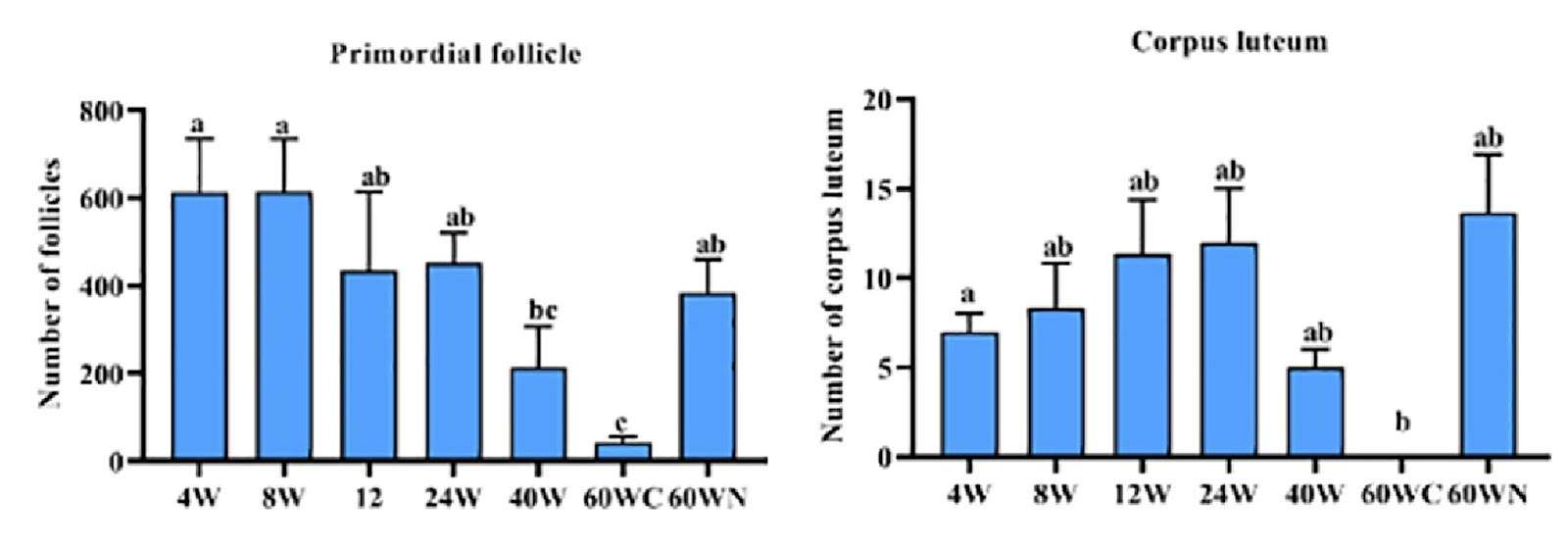
NMN administration enhances follicle development. The number of primordial follicles and corpus luteums span the ovarian cycle at different ages in female mice.
NMN decreases senescence marker in ovaries of mid-aged mice
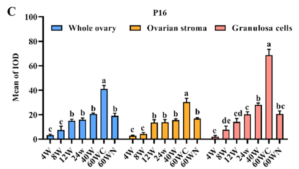
The p16 protein slows cell divisions and proliferation during the progression of senescence. Recently, elevated p16 levels have been considered one of the indicators of ovarian aging. The present study showed high levels of p16 in ovarian follicles at 60 weeks. Huang and colleagues found that NMN diminished the levels of p16 in mouse ovaries. The drop in the ovary’s levels of p16 suggests that NMN alleviates ovary tissue senescence and protects it during aging.
The Jiangsu University researchers then show that the improvement to ovarian senescence by NMN treatment is likely due to the upregulation of critical cellular processes. For example, Huang and colleagues show increased mitochondria function and the recycling of cellular damage and debris (autophagy) in the ovaries of mid-aged mice exposed to long-term administration of NMN. In addition, the protein balance and the antioxidant capacity of follicles were restored, thereby keeping follicles relatively young.
(Huang et al., 2021 | J Nutr Biochem.)
NMN administration decreases the levels of P16 protein. The protein level of p16 increases with age, reaching a peak in the 60-week control (60WC) group. Mice treated with NMN for 20 weeks beginning at 40 weeks of age (60WN) showed significantly decreased levels of p16.
These results indicate that the ovarian status of mice in the 60WN group was relatively younger after NMN supplementation.
Can a supplement a day keep ovarian aging at bay?
The importance of issues related to ovarian aging has been increasing progressively over the past several decades since, increasingly, more couples in all developed countries choose to postpone parenthood to more advanced female ages. This trend affects the likelihood of fertilization and conception, whether resulting from natural conception, conventional in vitro fertilization (IVF), or intracytoplasmic sperm injection (ICSI).
Since advanced male age seems to have little effect on the success rate for IVF/ICSI attempts using oocytes from young donors, much of the attention to improving the chances of parenthood has pointed to improving ovarian aging. Though treatment options need to be fine-tuned to reflect each woman’s condition, it is possible that supplementing with NMN can go a long way for improving the chances to experience some of life’s most miraculous adventures: pregnancy, childbirth, and parenthood.
References
Huang P, Zhou Y, Tang W, Ren C, Jiang A, Wang X, Qian X, Zhou Z, Gong A. Long-term treatment of Nicotinamide mononucleotide improved Age-related Diminished Ovary Reserve through enhancing the mitophagy level of granuloas cells in mice. J Nutr Biochem. 2021 Nov 18:108911. doi: 10.1016/j.jnutbio.2021.108911.