https://pubmed.ncbi.nlm.nih.gov/32744417/
First published: 03 August 2020 https://doi.org/10.1111/acel.13206
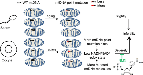
Mammals' aging is correlated with the accumulation of mitochondrial DNA (mtDNA) mutations.
Here, scientists analyzed oocyte (a cell in an ovary which may undergo meiotic division to form an ovum) quality of young (≤30 years old) and elder (≥38 years old) female patients and show the elder group had lower blastocyst formation rate and more mtDNA point mutations in oocytes.
Scientists show that mtDNA mutation levels inversely correlate with fertility, interestingly mainly affecting not male but female fertility. mtDNA mutations decrease female mice's fertility by reducing ovarian primordial and mature follicles. Scientists showed the mtDNA mutation types in oocytes during age.
Mechanistically, accumulation of mtDNA mutations decreases fertility by impairing oocyte's NADH/NAD+ redox state, which could be rescued by nicotinamide mononucleotide treatment.
For the first time, scientists answer the fundamental question of the causal effect of age‐accumulated mtDNA mutations on fertility and its sex dependence, and show its distinct metabolic controlling mechanism.
In short, NMN is viewed as a promising therapy for age‐associated physiological dysfunction and disease. We found NMN also has potential as a drug for mtDNA mutation caused oocyte aging.
In summary, our study, by systematically comparing the quality and mtDNA mutations of oocytes in young and elder female patients, showed that mtDNA point mutations inversely correlate with oocyte quality, which provides another potential biomarker for embryo viability in assisted reproduction, and demonstrated NMN as a potential candidate drug for oocyte aging caused by mtDNA mutation.
Aging is one of the key factors in both male fertility and female fertility. Indeed, female (human) fertility normally peaks at age 24 and diminishes after 30, with pregnancy occurring rarely after 50.
Mitochondrial malfunction has been hypothesized to play important roles in age‐ and environment‐induced infertility. For instance, mitochondrial DNA (mtDNA) deletions were reported to accumulate in human ovarian aging and mtDNA mutations may cause male infertility due to loss of spermatocytes and spermatids.
As a result, assessment of mitochondrial function status, mtDNA content, and mtDNA integrity is often performed to investigate the quality of sperms and oocytes in assisted reproductive technologies.
In humans, female fertility begins to decrease after the age of 30 and decreases more rapidly after 37.
Based on this, scientists divided female patients undergoing in vitro fertilization (IVF) or intracytoplasmic sperm injection (ICSI) into young (≤30 years) and elder (≥38 years) groups, and investigated the impact of age on oocyte mtDNA mutations.
What they found was that elder female patients of age ≥38 years have lower blastocyst formation rate and more mtDNA point mutations in oocytes than young female patients of age ≤30 years.
Fertilization rates and good‐quality embryo (2PN) rates showed no significant differences between young and elder, but blastocyst formation rate of elder group was significantly lower than that of young group in both IVF and ICSI cycles. This finding is consistent with a previous study showing that blastocyst formation declined with age.
Collectively, their results indicate that elder female patients have defects in blastocyst formation correlated with an increased accumulation of oocyte mtDNA point mutations.
Fertility 1
Accumulation of mtDNA mutations decrease female fertility by reducing oocyte's NADH/NAD+ redox state.
NADH/NAD+ has an important role in energy metabolism, and their redox state can be monitored in living cells using SoNar, a NADH/NAD+ sensor. These data indicate that oocytes have lower NADH/NAD+ redox ratio and weaker energy production.
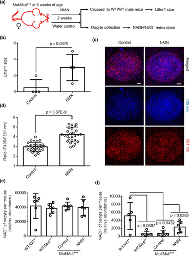
Accumulation of mtDNA mutations decrease female fertility by reducing oocyte's NADH/NAD+ redox state. NMN was added to subjects water for 2 weeks.
As female fertility is affected by aging in addition to the above‐mentioned mtDNA mutations, they further detected oocyte's NADH/NAD+ redox ratio in young and elder mice. They found a significantly decreased NADH/NAD+ ratio in elder mice as compared to the young counterparts. These results thus established a link between female infertility and perturbed NADH/NAD+ redox state.
As it has been reported that NMN is a promising therapy for aging‐associated physiological dysfunction and diseases through rescuing NADH/NAD+ redox state, they tested whether NMN could increase fertility of mice with high levels of mtDNA mutations.
As shown in Figure, the first litter size of female treated with NMN was higher than that from female with water.
This result indicates that NMN is remarkably capable of ameliorating infertility in female mice.
For female mice treated with NMN and water it was observed that the ratios in oocytes of mice with NMN were higher than mice with water, demonstrating an enhancement of the NADH/NAD+ ratio by NMN treatment.
Respective quantification of NADH and NAD+ revealed an increase in the amount of NADH, but not of NAD+, in oocytes of treated with NMN.
NMN treatment was shown to induce mitophagy in stem cells, leading to removal of dysfunctional mitochondria and thus cell function recovery.
Interestingly, NMN failed to alter the motility and ATP levels of sperm in male mice. All these results indicate that NMN can rescue fertility of PolgA mutator mice with more point mutations by enhancing cellular NADH/NAD+ ratio in oocytes.
For the first time, we quantified the effect of aging on the accumulation of heteroplasmic mtDNA mutations in human individual oocytes using next‐generation sequencing (NGS).
Aging of the human female reproductive system is much faster than that of other body systems, and follicle number reduction and oocyte quality decay with oxidative damage during ovarian aging cause the gradual decline in female fertility.
Scientists showed the mtDNA mutation types in oocytes during age.
This study also showed that mtDNA point mutations inversely correlate with oocyte quality, which provides another potential biomarker for embryo viability in assisted reproduction. Indeed, mitochondrial transfer has been used to exchange and enhance the integrity, activity, and number of mitochondria in quality‐compromised oocytes, which was recently used to improve fertility in women with previous poor reproductive performance by autologous mitochondrial injection treatment. This work provides a biomarker for the clinical application.
Our results answer the fundamental question—which step of oogenesis is damaged by age‐related mtDNA mutations—and suggested follicles could be the potential therapy target for female infertility. For sperm aging, mtDNA mutations may cause male infertility due to loss of spermatocytes and spermatids which can be rescued by increasing total mtDNA copy number.
However, our results demonstrate that mtDNA mutations specially reduce sperm motility without significantly compromising the fertility of young male. The levels of mtDNA mutations in oocytes versus other cell types, and the levels of heteroplasmy in offspring are a worthy follow‐up study, thus being a limitation of our present study.
NAD+/NADH redox state is known to play essential roles in cell metabolism. They demonstrated that mtDNA mutations decrease female POLG mutator mice's fertility by impairing oocyte's NADH/NAD+ redox state.
Interestingly, NAD+ availability was recently shown to decrease with age. Hence, this study further emphasizes important roles of NADH/NAD+ redox state in oocyte aging. The perturbed NADH/NAD+ redox state may further compromise energy metabolism, as is observed for the oocytes of POLG mutator mice.
NMN, a key NAD+ intermediate, has been shown to enhance NAD+ biosynthesis, activate SIRT1, and improve metabolic and stress responses in aging mice as well as ameliorate various pathologies in mouse disease models.
Consistently, NMN treatment elevated the amount of NADH, the reduced form of NAD+, in oocytes of POLG mutator mice. The unchanged amount of NAD+ with the up‐regulation of NADH indicated that the overall size of NADH/NAD+ pool in oocytes was increased by NMN treatment.
Fertility 2
Thus, NMN can be considered as a potential agent in the treatment of cell metabolism disorders triggered by perturbed the overall of NADH/NAD+ pools.
NMN supplementation has been reported to reverse age‐related arterial, vascular, and skeletal muscle dysfunction in mice by mitochondrial‐related signaling. Indeed, it was shown to be transported into mammalian mitochondria.
The short‐term administration of NMN has been reported to have remarkable therapeutic effects on metabolic complications and other disease conditions.
In short, NMN is viewed as a promising therapy for age‐associated physiological dysfunction and disease. We found NMN also has potential as a drug for mtDNA mutation caused oocyte aging. Further mechanistic studies are required in future.
In summary, our study, by systematically comparing the quality and mtDNA mutations of oocytes in young and elder female patients, showed that mtDNA point mutations inversely correlate with oocyte quality, which provides another potential biomarker for embryo viability in assisted reproduction, and demonstrated NMN as a potential candidate drug for oocyte aging caused by mtDNA mutation.
Water consumption was measured for 2 weeks prior to the start of NMN administration. NMN was administered in drinking water at 900 mg/kg/day, based on the previously measured water consumption (Mills et al., 2016).
The administration began at 6 weeks of age and continued for 2 weeks. The NMN solution was prepared weekly in small batches by dissolving NMN into autoclaved water at the certain dose and filtering sterilely. Water bottles and cages were changed twice weekly.